
Why Is The Inside Of My Ear Itchy? The Causes Behind The Tickle You Can’t Reach
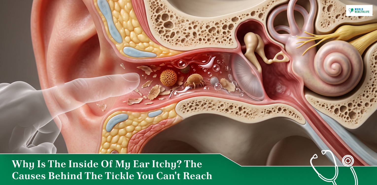
Few itches are as maddening as the one you physically cannot scratch. If you keep wondering “why is the inside of my ear itchy?” then you are dealing with one of the most common complaints in ear care. Usually, this has a simple explanation. Moreover, the usual suspects are dry skin, trapped moisture, and an allergy. Occasionally, though, the itch is an early sign of an infection or a skin condition that needs treatment. So, the following guide will explain what is happening in the canal, the most likely causes, what to do at home, and when to see a doctor. Why Is The Inside Of My Ear Itchy? How The Ear Canal Protects Itself First, let us understand the overall biology of your ear canal. The ear canal is a narrow tube lined with thin skin, which is packed with nerve endings. Moreover, it is responsible for producing earwax. The primary function of earwax is to trap dust and keep the canal slightly acidic. In this way, bacteria and fungi struggle to grow. When this system is thrown off, the nerves react with itching before any other symptom shows up. That is why a small change in moisture or wax can feel so irritating. Wax, Moisture, And The Skin Barrier Earwax, medically termed cerumen, acts as a natural moisturizer and shield. If you remove too much of it, the skin starts to dry, flake and itch. Conversely, too much moisture from sweat, showers, or swimming leads to the skin softening. As a result, this allows microbes to settle in. So, the itch comes from either extreme. Why Is The Inside Of My Ear Itchy But Not Painful? Itch and pain travel along different nerve fibers. Early irritation, allergy, and dryness trigger itch signals first, while pain usually appears once swelling or infection builds. So, an itchy ear with no pain is often an early or mild problem. Still, it can become painful within a day or two if the cause is infectious. Thus, watch the way it changes. Common Causes Of An Itchy Ear Canal Most cases trace back to a handful of causes. The following breakdown evaluates on how you can tell them apart, along with the sections CauseTypical CluesUsually Goes Away?Dry skin or too little earwaxFlaky skin, mild itch, no dischargeYes, with gentle careSwimmer's ear (otitis externa)Itch, then pain when you tug the ear, possible dischargeYes, with prescription ear dropsFungal infection (otomycosis)Intense itch, fullness, white or black debrisYes, with targeted antifungal treatmentEczema or seborrheic dermatitisRed, scaly skin, itch elsewhere on face or scalpControlled, but it frequently recursAllergiesItchy ears with sneezing, runny nose, itchy eyesYes, once the primary trigger is treatedHearing aids or earbudsItch occurs only when the device is in useYes, with a better fit or different materialEarwax buildupSensation of fullness, muffled hearing, itchYes, after safe and proper removal Dry Skin And Over-Cleaning When you use cotton swabs, hairpins, and fingernails to clean your ears, it strips away the protective wax while scratching the delicate lining. Following this, the skin becomes dry and itchy, and scratching makes it even worse Many people get stuck in this loop because the itch drives them to clean more. However, leaving the canal alone usually solves the problem within a week or so. Swimmer’s Ear And Fungal Infections When you have trapped water in your ears, that is generally a classic trigger. Bacteria cause swimmer’s ear, and it often starts as itching before turning painful. On the other hand, fungi cause otomycosis. This is more common in humid climates and in people who use hearing aids or have used antibiotic drops for a long time. A fungal infection tends to itch intensely and may leave fluffy white or dark specks in the canal. However, remember one thing: neither of these responds to home remedies. So, you need the right drops. Eczema, Dermatitis And Psoriasis Skin conditions can affect the ear canal just as they do the rest of the body. For instance, seborrheic dermatitis shows up as greasy flakes, often along with dandruff. Eczema can make the canal red and scaly. On the other hand, when you have contact dermatitis caused by shampoo, hair dye, or nickel in earrings and earbuds is also common. If your ears itch alongside a rash on the scalp or behind the ears, a skin condition is likely. Allergies And The Eustachian Tube Hay fever and food allergies can cause itching deep in the ear, often described as an itch you can feel but not reach. This happens because the eustachian tube, which connects the ear to the back of the throat, becomes inflamed. Some people with pollen allergies also feel itchy ears after eating raw fruit or vegetables, a reaction known as oral allergy syndrome. Antihistamines or allergy nasal sprays often calm it. Itchy Ears With Other Symptoms The symptoms that usually follow the itching tell you a lot. When you have an itch only, it is usually something minor. However, additional symptoms help narrow the cause and show how urgent it is. Symptom with the ItchWhat It May SuggestWhat to DoSneezing, runny nose, itchy eyesAllergyTry an over-the-counter antihistamine and avoid known triggers.Pain when pulling the earlobeSwimmer's ear (otitis externa)See a doctor for prescription antibacterial ear drops.Discharge or unusual odorOuter or middle ear infectionBook a medical appointment for evaluation and proper treatment.Muffled hearing or fullnessEarwax buildup or fluid behind the eardrumGet the ear examined by a healthcare professional for safe removal.Itchy, flaky scalp and faceSeborrheic dermatitis or eczemaUse targeted medicated shampoo and ask a doctor about ear-safe creams.Dizziness or ringing (tinnitus)Inner ear problemSee a doctor promptly for a comprehensive neurological and ear exam. What You Can Do At Home For mild itch with no pain, discharge, or hearing change, gentle care is enough. The goal is to protect the skin and keep the canal dry without digging into it. Moreover, a few habits make most of the difference. Target Area / ActionSafe Steps (Do This)What to Avoid (Don't Do)Daily CleaningClean only the outer ear with a damp cloth. Leave the ear canal completely alone.Never insert cotton swabs, keys, or pen caps into the canal. They scratch skin and push wax deeper.Moisture ManagementDry ears after swimming/showering by tilting your head and using a towel or a cool, arm's-length hair dryer.Do not scratch wet or itchy ears. Even a small fingernail scratch can introduce a painful infection.Water ActivitiesUse earplugs or a swim cap if you are highly prone to developing swimmer's ear.Do not ignore trapped water or let moisture sit in the canal for extended periods.Devices & WearablesClean hearing aids and earbuds regularly and give your ears frequent breaks from using them.Do not leave dirty devices in your ears, as they trap moisture and introduce harmful bacteria.Home RemediesTreat underlying allergies. Controlling hay fever symptoms often settles the phantom ear itch automatically.Avoid ear candles and unverified oils (like garlic or peroxide). They can burn skin or damage a perforated eardrum. How Doctors Treat An Itchy Ear Your treatment is dependent on the cause. So, the doctor will first look into the canal with a small lighted scope. That quick exam usually reveals wax, flaking skin, or signs of infection. From there, the options are straightforward. Antibiotic or antifungal ear drops treat infections. Moreover, some contain a steroid to reduce swelling. Steroid drops or creams can further help seborrheic dermatitis. However, if wax is the problem, a clinician can remove it with gentle suction or a small instrument. Allergies are treated with antihistamines, nasal sprays, or allergy testing when symptoms keep returning. When To See A Doctor Most itchy ears settle within a few days once you stop irritating them. Book an appointment if the itch lasts more than a week, keeps coming back, or you can't stop scratching. See a doctor sooner if you notice pain, discharge, swelling, a bad smell, or any drop in hearing. Get prompt care if the itch comes with fever, swelling behind the ear, severe pain, dizziness, or weakness in the face. These can signal a deeper infection. People with diabetes or a weak immune system should be seen early, because ear infections can spread more easily in these cases. An itchy ear is rarely dangerous, but it is rarely random either. Keep the canal dry, resist the urge to dig, and pay attention to what comes with the itch. If it won't go away, a quick look from a clinician is usually all it takes to end it. Disclaimer: The information provided in this article is for general informational purposes only. It does not, and is not intended to, constitute medical or health advice. Please consult a qualified healthcare professional for medical guidance or expert health assistance. References Cedars-Sinai. (2025). Is It Really Dangerous to Clean My Ears with Cotton Swabs? Cedars-Sinai. Centers for Disease Control and Prevention. (2026). Preventing swimmer’s ear. Healthy Swimming. Centers for Disease Control and Prevention. Lipschutz, J. (2024). What Is Earwax? Understanding Its Role and Importance. Berkeley Hearing Center. Eimers Hearing Clinic. (2026). The risks of Over-Cleaning your ears. Dr. Eimers Hearing Clinic. Mitchell, K., & Gatta, F. (2026, June 17). How to clean your ears. WebMD.


Browse By Category


LATEST NEWS




Living Healthy


OUR BLOG SPACE
View AllHow Long Is Too Long in the Wrong Job Before Your Health Suffers?
How long should you stay in a job? Most people may never say it out loud, but many are trapped in a job that brings them nothing but stress. It’s not too unprofessional to say it as it is: “I hate my job,” let alone switch too soon. This is where things get interesting: who decides when it’s too soon, or when it's too long in the wrong job? Some believe that waiting even six months can leave a permanent dent in one’s mental health. Others try to drag the wagon until they can't anymore. As for traditional career advice, it isn’t any better with its caution against leaving before two years to avoid looking like a job hopper. For perspective, Gallup’s State of the Global Workplace 2026 report found that 40% of employees worldwide experienced significant stress the day before the survey. It’s not just about what you’re sacrificing in the current moment. The repercussions often stretch out, especially when workplace stress makes it harder to rest and recover. Not every difficult job is the wrong one, and leaving does involve a different set of challenges. How do you know when perseverance is costing you too much? This article explores how prolonged workplace stress can affect your well-being and when it may be worth reconsidering your circumstances. The Deepening Roots Of Pressure And Stress Anybody who works for a living knows that pressure is the name of the game, at least to an extent. You might encounter an unusually challenging client or a deadline that just won’t let you sleep. Once in a while, that’s not a major issue. The question is whether your mind and body can bounce back to equilibrium. If respite feels strange, there’s a problem. In such cases, even formal leaves or weekends can feel restless, with a gnawing anxiety masking the rest. In its 2026 Work in America survey, the American Psychological Association (APA) found that 95% of US workers wanted to work for an organisation that valued their emotional well-being. Still, only 80% described their workplace as psychologically healthy. A clear disconnect exists between what people value and what they ultimately experience. Take comfort in knowing you’re never truly alone in your experiences. Start by paying attention to the following signs, as they indicate that the root of work-related stress is getting deeper and stronger: You can't shake off a nagging exhaustion, even after a night's sleep or a quiet weekend. Work-related worries won’t allow you to stay asleep, even forcing you to wake up through the night. Even the smallest inconveniences feel like a mountain you don’t want to deal with. It’s a whole new task to stay focused and remember things that would usually come easily. You’re complaining of frequent headaches, muscle tension, and stomach problems. Even life outside of work can feel like drudgery, whether it's hobbies or social plans. Stress can have many causes, but if you see these signs, don’t take them lightly. If it's been weeks or months since you felt joy and enthusiasm for your work, it’s time to pause. How Long Should You Stay In A Job: Cost Of Staying Vs. Cost Of Leaving Most people who stay in a highly stressful job have legitimate reasons, mainly responsibilities. Their income supports their household, or they may have spent years building their career. When so much is at stake, it becomes natural to tell yourself things will improve in due time. Truth be told, sometimes the tide does turn for the better. However, if the same pressures remain unresolved, it makes little sense to put your work life on waiting mode. Does that mean you lean toward the other extreme, leaving? Not until you’ve weighed the cost of staying against the cost of leaving. The table below can give more perspective on this: Decision PathImmediate Reality & BenefitsHidden Costs & Long-Term RisksStaying PutYou lock in a completely predictable routine and a dependable paycheck. Plus, you can leverage your track record to negotiate a better deal right where you are.The grinding, unresolved friction will slowly drain your emotional battery. You skip immediate drama, but the toxic roots stay exactly the same.Jumping ShipBreaking free gives you a blank canvas for a total career rebirth. You will aggressively level up your skills and open doors to much bigger opportunities.The job hunt itself is an exhausting, short-term uphill battle. The transition demands hard cash and time, and the new office will come with its own messy pitfalls. Testing New Waters Without Jumping Ship Some people view this period as a chance to move towards work that better reflects their interests or values. So, how long should you stay in a job? For instance, someone drawn to caring for others might explore becoming a nurse online instead of immediately stepping away from existing responsibilities. Online study can make that exploration more manageable for someone balancing employment, family commitments, and education. Elmhurst University notes that with a computer and a stable internet connection, you can complete much of the coursework from home, a library, or even a cosy coffee shop. That being said, no field is completely free from stressful tasks. So, ask yourself whether the change you’re considering would bring you closer to the kind of work you want. Before coming to a decision, check both sides of the scale. A Passing Season Or A Desperate Plea For A Change? Has it been a long time since you told yourself, ‘this too shall pass’? Perhaps you have no concrete answer. When work affects one’s well-being, it can be difficult to make sense of things. You may wonder whether this is just another rough patch or a problem that won’t resolve on its own. The answer lies in the pattern, not a single bad day. The following three scenarios can help you understand what your experience might be communicating: A Clear End To The Pressure Perhaps your team is handling an unusually demanding project or a colleague is on leave. That generally increases others’ workload. You could feel utterly exhausted and distracted. The silver lining is that you know what’s causing the pressure and that it will likely ease in due time. One solution is to figure out whether things can be made manageable for now. Discuss priorities with your manager or ask for support where you need it. Then, once the demanding period has passed, you can reevaluate if things have changed or returned to normal. A realistic endpoint to work-related stress or pressure is usually not bad. It simply means you’re facing a difficult season that shall truly pass. Every Week Feels The Same, Full Of Monotony Does week after week look painfully the same? It’s gotten so challenging that you dread even Sunday evenings or vacations. Worse, you keep promising yourself the next week will be different, only for the same cycle to play out again. The longer this pattern continues, the harder it can feel to imagine a way out. According to a recent Fortune report that cited a Glassdoor poll of over 1,300 professionals, 53% of US workers surveyed had paused their job search. Why? So they could protect their mental health. Well, they may have noticed the unmistakable signs. Jade Walters, a TEDx speaker and founder of The Ninth Semester, put it bluntly: “One of the biggest signs of exhaustion is noticing a lack of emotional regulation. You’re more irritable, more anxious, and more frustrated.” That’s when even looking for a new, better job feels like a chore not worth the time and effort. The Work Is Great, The Workplace Not So Much In many cases, the problem is not the profession itself. You may find genuine enthusiasm and joy in your work but struggle with a manager who offers little support or fosters a culture of micromanagement. Then, a different team, schedule, or role may become a better option. If changing your circumstances internally is realistic and safe, explore that possibility. If the same issues run throughout the organisation, it may make more sense to look elsewhere. Protect Your Health, Whether That Means Starting Over Or Otherwise After a long discussion, let’s address the million-dollar question: How long is too long in the wrong job? The truth is that there is no universal answer. What one person may slide by during a difficult season, another may find unsustainable. It all depends on one’s responsibilities, support system, and personal well-being. The National Alliance on Mental Illness (NAMI) revealed in a 2026 poll that 26% of the surveyed US employees said they had considered quitting because of mental health impact. Financial commitments, family responsibilities, and uncertainty about the future made the decision difficult. If your situation is similar, leaving all at once may be out of the question, and that’s fine. A gentler transition may be required, one that involves making smaller changes first. Start with the steps that feel manageable, as follows: But how long should you stay in a job? Draw a hard line in the sand once, then defend it. You could ghost your notifications at five or isolate your lunch hour from the creep of spreadsheets. Lay out the gritty details of your crushing workload to your manager and demand a concrete change. Lock down your calendar: Prioritise deep sleep, physical movement, and the relationships anchoring you to reality. Activate support networks: Lean on a trusted confidant or tap workplace resources before you hit a total psychological wall. Keep your escape hatches well-greased by sprucing up your resume. If cutting ties is your only path to survival, calculate your financial runway with cold precision. Watch what happens next. If dysfunction remains stubbornly unchanged, you will have the clarity needed to make your next big move.
Why Are My Armpits Itchy? A Deep Dive Into Underarm Irritation
Having a persistent itch in your underarms can not only be an incredibly frustrating experience but also highly uncomfortable. The underarm area is naturally sensitive, prone to moisture, and subjected to daily friction. So, it is highly vulnerable to various forms of irritation. Thus, if you are constantly asking yourself, “why are my armpits itchy?”, you are not alone. Having discomfort in your underarms is so common that it can stem from the following: Everyday lifestyle choices Environmental factors Underlying dermatological conditions. Therefore, having an understanding of the primary causes of this irritation is the first step toward finding lasting relief. Below, we dive deep into the most frequent triggers behind itchy armpits and look at the red flags that warrant a doctor’s visit. Besides that, we will also outline actionable steps you can take immediately to soothe compromised skin. The Primary Triggers Of Underarm Itching Think about how thin the skin is inside your axillary vault. It folds tightly against itself all day. Plus, it is packed densely with sweat glands. This specific biological layout makes the whole area uniquely vulnerable to severe irritation. Disrupt this localized ecosystem even a little, and your body hits defense mode fast. A sharp inflammatory response flares up. That reaction is what registers as a persistent, maddening itch. To make it stop, zero in on the exact factors attacking your skin barrier. It could be chemical, environmental, or biological. At A Glance Primary TriggerCommon CauseKey SymptomsChemical Contact DermatitisDeodorants, fragrances, baking sodaLocalised redness, burning, irritationIntertrigo & Fungal GrowthTrapped moisture, skin friction, yeastBright red, raw, scaly rashMechanical DamageShaving, dull razor blades, frictionRazor burn, small red bumps, pimplesSystemic ConditionsEczema, psoriasis, immune issuesChronic itching, deep lumps, dry patches Chemical Contact Dermatitis: Disruption By Deodorant One of the most frequent reasons people experience underarm itching is contact dermatitis. This occurs when the skin reacts negatively to a foreign substance. There are two main types here. TypesDescriptionIrritant Contact DermatitisA direct irritation caused by harsh ingredients that strip the skin's protective barrierAllergic Contact DermatitisA specific immune response to an allergen present in your personal care products Here, the daily deodorant that you use is the primary culprit. Moreover, a variety of traditional products contain the following: Heavy synthetic fragrances Alcohol, preservatives, such as parabens Baking soda. All of these can severely disrupt the delicate skin barrier. Laundry detergents and heavily scented body washes can leave behind residues on your clothes or skin. This further triggers an aggressive and itchy response. Intertrigo And Fungal Growth: The Warm-Zone Trap If you had to describe the human armpit, it is a naturally warm, dark, and humid environment. So, when skin rubs against skin, the result of this is friction and trapped sweat. This can lead to an inflammatory condition known as intertrigo. A further byproduct of this is an inflammatory condition known as intertrigo. During exercise or in hot weather, this is especially inevitable. But if it is ignored and left unmanaged, this moist microclimate becomes an ideal breeding ground for microscopic organisms. The most common culprit is a yeast called Candida. Typically, an armpit yeast infection causes a bright red, intensely itchy, sometimes scaly rash. In some cases, it may also feature small, pimple-like bumps (satellite lesions) spreading from the main irritated area. Mechanical Damage: Razor Burn And Folliculitis Shaving is another highly invasive routine for the underarms area. However, shaving with a dull blade is even worse. Dragging it across your skin or shaving it too closely ultimately leads to the creation of micro-tears in the skin barrier. Therefore, this mechanical friction causes razor burn. Generally, this presents as immediate stinging, redness, and a sharp itch as the skin attempts to heal. Furthermore, these micro-tears allow surface bacteria to enter the hair follicles. This can result in folliculitis. This is actually an infection or inflammation of the hair follicles that looks like small red bumps or white-headed pimples centered around underarm hairs. Folliculitis can be deeply itchy, tender, and prone to further irritation if it is shaved again. Systemic And Immune-Mediated Skin Conditions Sometimes, the root cause is not external. Instead, chronic skin conditions frequently target body folds like the underarms. ConditionCore MechanismKey SymptomsAtopic Dermatitis (Eczema)Genetic variation that weakens the skin's protective barrierIntense itching, severe dryness, and weeping patchesInverse PsoriasisInflammatory condition thriving specifically in body foldsSmooth, red, raw-looking patches that itch and burnHidradenitis SuppurativaChronic blockage of the underarm hair folliclesPainful, deep pea-sized lumps that can rupture Red Flags: When The Itch Signals Something Serious Most cases of underarm discomfort stem from superficial skin irritation. However, persistent or unusual symptoms sometimes warrant closer investigation. Chronic, unexplained underarm itching can occasionally serve as an early indicator of internal metabolic issues such as diabetes. This occurs due to blood sugar imbalances, triggering localized nerve irritation and systemic dry skin. Moreover, this often manifests as a stubborn, deep-seated itch in body folds. On rare occasions, chronic axillary irritation can signal oncological conditions. Though this connection is highly unusual, specific systemic signs require a prompt medical evaluation if they accompany a persistent underarm itch. These critical warning indicators include hard, painless, swollen lymph nodes in the armpit, drenching night sweats, or rapid unexplained weight loss. Thus, try seeking professional medical guidance for these symptoms. That is because it is essential to rule out rare but serious underlying conditions. Warning Signs: In A Nutshell Warning SignPotential Internal CauseKey Medical SignalChronic Axillary ItchingDiabetes / Metabolic IssuesLocalized nerve irritation and systemic dry skinHard, Swollen Lymph NodesHodgkin LymphomaOncological trigger affecting the lymphatic systemDrenching Night SweatsSystemic IllnessInternal regulatory dysfunction requiring prompt screeningUnexplained Weight LossAdvanced Systemic DiseaseRapid metabolic changes not linked to diet or exerciseSkin Thickening or DimplingInflammatory Breast CancerRare oncological link causing localized tissue changes What To Do Immediately If Your Armpits Won't Stop Itching? When underarm irritation gets this bad, you have to treat the skin like an open wound. That means completely freezing your daily grooming routine. Putting harsh chemicals on your skin or running a razor over raw, damaged areas drives the inflammation deeper and risks a nasty secondary infection. Your absolute first step is to cut out every single potential trigger. Stop using all deodorants, antiperspirants, and body washes immediately. This is especially if they contain heavy perfumes or baking soda. Next, throw on some loose, breathable cotton clothes. This keeps the area dry and stops your skin from constantly rubbing against itself. To take the edge off that non-stop, agonizing urge to scratch, press a cold compress against the area for 10 minutes at a time. You can also put on a thin layer of over-the-counter hydrocortisone cream or calamine lotion to help calm down the local nerves while your skin barrier tries to heal. Skip the home remedies entirely and get a doctor to look at it if the itching comes with any of these: Oozing sores A foul smell Extreme swelling A fiery red rash that’s actively spreading down your torso You also need to see a doctor. This means you experience an intense itch alongside deep, painful lumps under the skin, a sudden fever, or night sweats. Those are clear signs that you're dealing with something much deeper than a simple surface irritation. Disclaimer: The information provided in this article is for general informational purposes only. It does not, and is not intended to, constitute medical or health advice. Please consult a qualified healthcare professional for medical guidance or expert health assistance.
Searching “Why Is My Throat Itchy?” Know The Common Causes And Treatment Strategies
An itchy throat can be quite an annoying yet worrying thing to have. This persistent tickle often leaves people wondering, “why is my throat itchy,” as they scramble for immediate relief. In most cases, that irritating, scratchy sensation is a common and non-serious response to everyday environmental triggers or minor illnesses. Environmental factors such as seasonal allergies, dry indoor air, and airborne irritants such as tobacco smoke are frequent culprits that inflame the sensitive lining of the pharynx. Alternatively, an itchy throat can serve as the earliest warning sign os an impending viral infection, such as the common cold flu or viral pharyngitis. Less frequently, it may point to chronic underlying conditions such as gastroesophageal reflux disease (GERD) or even specific food allergies. Pinpointing the exact cause of your discomfort is the essential first step toward finding targeted, lasting relief and restoring your overall peace of mind. Causes Of An Itchy Throat That irritating tickle in your throat is rarely a standalone condition. Instead, it is a localized symptom triggered by an underlying disturbance in your body. When the sensitive nerve endings embedded in the mucosal lining of your pharynx become irritated, inflamed, or dried out, one thing happens. They send distress signals to your brain that result in the urge to cough or scratch. Pinpointing the exact source of this discomfort is crucial, as the root cause can range from simple environmental changes to viral infections or chronic digestive issues. Root CauseKey Identifying SymptomsAllergiesClear runny nose, sneezing, itchy eyes, seasonal patternsInfectionsFever, body aches, swollen glands, raw sore throatIrritantsSudden onset after exposure to smoke, pollution, or chemical fumesDry Air / DehydrationMorning throat tightness, dry mouth, chapped lipsPostnasal DripFrequent throat clearing, nighttime coughing, mucus sensationAcid RefluxHeartburn, sour taste in mouth, hoarseness, chronic dry cough Allergies Allergic rhinitis, commonly known as seasonal or perennial allergies, is one of the most frequent drivers of a persistently itchy throat. When you inhale harmless environmental particles such as pollen, dust mites, mold spores, or pet dander, an overactive immune system misidentifies them as dangerous invaders. In response, your body floods your system with histamines, a chemical compound that induces localized inflammation, swelling, and severe itching along the respiratory tract. This reaction typically presents alongside watery eyes, sneezing, and a clear, runny nose. Infections An itchy throat is frequently the earliest warning sign that a viral or bacterial infection is taking hold in your upper respiratory tract. In the initial stages of the common cold, influenza, or viral pharyngitis, replicating pathogens irritate the surface cells of the pharynx before full-blown pain develops. If the culprit is bacterial, such as strep throat, the itching will rapidly escalate into severe pain when swallowing. This is often accompanied by a sudden fever, body aches, fatigue, and visibly swollen lymph nodes in the neck. Irritants Your throat lining is highly sensitive to the air you breathe, where airborne physical or chemical irritants can cause immediate localized inflammation. Inhaling secondhand tobacco smoke, vaping aerosols, smog, industrial air pollution, or harsh household cleaning chemicals directly strips away the throat’s protective moisture barrier. This exposure causes mechanical and chemical irritation of the nerve endings. As a result, it causes a dry and scratchy tickle that usually subsides once you move into a clean and well-ventilated environment. Dry Air Or Dehydration When the ambient air lacks moisture, or when you fail to drink enough fluids, the mucus membranes lining your throat quickly dry out. This is particularly common during winter months when indoor heating systems sap Postnasal Drip Postnasal drip occurs when your body produces excess, thickened mucus that continuously trickles down the back of your throat instead of draining normally through the nose. This chronic dripping can be caused by sinus infections, allergies, or changes in the weather. As the mucus continuously washes over the sensitive tissues of the lower pharynx, it acts as a constant physical irritant. Furthermore, it triggers a chronic throat tickle, an involuntary nighttime cough, and a constant, annoying urge to clear your throat. Acid Reflux Gastroesophageal Reflux Disease (GERD) or silent reflux (laryngopharyngeal reflux) occurs when the muscular sphincter at the top of your stomach fails to close properly. As a result, this lets harsh gastric acid travel up the esophagus. And when these acidic stomach contents reach the delicate tissues of the throat, they cause micro-burns and chronic chemical irritation. Thus, because this tissue lacks the protective lining found in the stomach, even trace amounts of acid do the job. That is, a persistent, unexplained throat tickle, hoarseness, a sour taste, and a dry cough. Simple Ways To Find Relief For An Itchy Throat Treating an itchy throat relies on two main approaches. They are: delivering immediate moisture to soothe irritated nerve endings addressing the underlying inflammation This is fully dependent on what is triggering your discomfort. So, you can find effective relief using common household ingredients or targeted over-the-counter remedies. Immediate Home Remedies When a sudden tickle strikes, your kitchen cabinet is often the best first line of defense. Simple, natural ingredients can coat the throat and ease discomfort without the side effects of medications. These home treatments focus on the rehydration of dried mucosal membranes, calming overactive nerve endings, and reducing the physical friction caused by swallowing and coughing. Targeted Over-The-Counter Options When natural home remedies are not enough, turning to pharmacy shelves can provide fast, clinically targeted relief. Over-the-counter medications work by directly interrupting the biological triggers of your discomfort. That means physically blocking allergic histamines, numbing overstimulated nerve endings, or clearing nasal congestion that feeds a throat tickle. Choosing the right product allows you to address the specific root cause of your symptoms. At A Glance Remedy TypeSpecific TreatmentHow It WorksQuick Tips / PrecautionsImmediate Home RemediesWarm Saltwater GarglesDraws excess fluid out of inflamed tissues to reduce swelling; washes away trapped allergens or mucus.Mix ½ tsp salt into 8 oz warm water. Gargle for 30 seconds several times a day.Raw HoneyActs as a natural hypertonic demulcent to form a thick, protective physical barrier and soothe inflammation.Take 1–2 tsp directly or mix into warm water/tea. Never give to infants under 1 year.Steam Inhalation & HumidifiersDirect inhalation of moist air lubricates dry passages, thins stubborn postnasal drip, and stops dry-air irritation.Use a cool-mist humidifier in the bedroom or breathe in steam from a hot shower.Aggressive Fluid IntakeConstant hydration thins mucus secretions and prevents the physical friction that triggers a dry cough.Sip 8–10 glasses daily of warm broths, diluted warm apple juice, or decaf herbal teas.Targeted OTC OptionsDemulcent LozengesStimulates saliva flow to coat the throat. Variants with menthol or benzocaine temporarily numb sensitive nerve endings.Look for ingredients like pectin, glycerin, or honey.OTC AntihistaminesHalts the systemic allergic response by blocking histamines at the receptor level.Best if the itch is caused by allergies. Examples: cetirizine, loratadine, fexofenadine.Saline Nasal Sprays & RinsesFlushes out accumulated pollen, dust, and thick mucus before it can travel down the throat as postnasal drip.Use a saline nasal spray or a neti pot. When To Consult A Healthcare Professional Most cases of a scratchy throat resolve on their own. However, there are certain signs that indicate it is time to seek professional care. So, you need to consult a healthcare professional if your symptoms persist for more than a week without improvement. In addition to that, seek care if your itchiness rapidly escalates into a severe sore throat. Do the same if you experience a high fever, chills, or difficulty swallowing. Also, get evaluated if you have unexplained hoarseness or voice changes lasting over two weeks. This issue can point to chronic acid reflux or vocal cord strain. On a final note, seek immediate emergency medical attention if your throat tickle is accompanied by severe red flags. This includes difficulty breathing, wheezing, structural swelling of the neck or tongue, or an inability to manage your own saliva. Disclaimer: The information provided in this article is for general informational purposes only. It does not, and is not intended to, constitute medical or health advice. Please consult a qualified healthcare professional for medical guidance or expert health assistance. References Cleveland Clinic. (2025). Itchy throat. Cleveland Clinic. Ascent ENT Hospital. (2025). Why Is My Throat Itchy? Common Causes, Symptoms, and Relief. Ascent ENT Hospital. Fletcher, J. (2025). What to know about itchy throat causes, treatments, and related symptoms. Medical News Today.
Wondering “Why Is My Vagina Itchy?” Know The Causes, Remedies And When To Consult A Doctor
When it comes to proper vaginal health, hygiene is of utmost importance. Moreover, the most important part of that is maintaining its pH. However, any disruptions to that health, be it an itch or a burning sensation, can give any lady quite a scare! But here is the thing. Vaginal itching is actually quite a common phenomenon, where it affects approximately 75% of women at some point in their lives. As a result, it is one of the most common feminine health concerns. So, if you find yourself anxiously and quite irritatingly wondering, “why is my vagina itchy?” do not worry. The good news is that most vaginal itching cases resolve within a week of home treatment. However, persistent symptoms lasting beyond this period may require you to warrant a doctor’s evaluation. Thus, considering everything, the following guide aims to give you a detailed breakdown. What Is Vaginal Itching Before we understand the science behind an itchy vagina, first identify the area of irritation. Identify The Area Of Itching If the itching occurs in your visible genital area, including your clitoris, vulva, and pubic area, then what you are facing is vulvitis. This is vulvar itching. Conversely, if the itching is in your vagina, which is the internal muscular canal leading up to the cervix, then it is vaginal itching. A Breakdown Of Vaginitis First, let us break down the term. Medically, this is termed vaginitis. The term literally translates to inflammation of the vagina, where vagin is vagina and it is inflammation. In vaginitis, there is inflammation or irritation of the vagina and the outer genital area, the vulva. This frequently causes vaginal itching, redness, soreness, pain, or abnormal discharge. The literal breakdown means inflammation. In modern medicine, vaginitis is used as an umbrella term for a variety of conditions that cause irritation, abnormal discharge, itching, or pain in the vaginal area. It is most commonly triggered by an imbalance in the natural vaginal flora, microbial infections (such as yeast or bacteria), chemical irritants, or hormonal changes. Vaginal Itching Vs. Vulvar Itching The primary difference between vulvar and vaginal itching comes down to location and root cause. The vulva is the outer, visible genital area, which includes the lips and your clitoris. Conversely, the vagina is the internal canal. Thus, itching in these areas differs. Point of DifferenceVulvar ItchingVaginal ItchingActual Term UsedPruritus Vulvae / VulvitisVaginitisLocation The external skin and folds (labia majora, labia minora, clitoris, and vaginal opening)Inside the internal muscular canal leading up to the cervixCommon Causes• Contact dermatitis • Skin conditions such as eczema, psoriasis, lichen planus, etc.• Frictional or environmental factors • Hormonal changes• Infections such as yeast infections • Yeast infection or candidiasis• Bacterial Vaginosis (BV)• Sexually Transmitted Diseases (STDs)• Hormonal shifts such as fluctuations in estrogen altering the vaginal lining and pHTypical Symptoms • Visible redness• Swelling• Skin cracks• Raw patches• Burning• Soreness • Internal itching or irritation• Changes in vaginal discharge (color, consistency, or smell)• Sometimes discomfort or pain during urination or sex Common Causes Behind An Itchy Vagina An itchy vagina may be common and highly treatable. However, you still need to understand the wide range of factors that actually contribute to yours. Understanding the distinct mechanisms and secondary symptoms behind each cause helps pinpoint the root issue. Yeast Infections A vaginal yeast infection occurs when there is an overgrowth of Candida, a fungus that naturally lives in the vaginal ecosystem in small numbers. The vagina is typically kept in check by beneficial bacteria known as Lactobacilli. However, when this balance is disrupted, the yeast tends to thrive and trigger inflammation. The key accompanying symptoms include having an intense itching and burning in the vulva and vagina. Moreover, it can cause thick, odorless white discharge that resembles cottage cheese. Apart from these, there is also redness, swelling, and superficial micro-cracks in the skin. Bacterial Vaginosis (BV) Bacterial vaginosis (BV) is considered the most common vaginal infection for women of reproductive age. Though linked to sexual activity, the condition is not an STI. BV arises when the natural vaginal flora shifts, which causes a significant drop in protective lactobacilli and a massive overgrowth of anaerobic bacteria. This alters the vaginal pH, which shifts it from healthy acidity to alkalinity. Consequently, this creates localized irritation. Along with mild to moderate itching, if you have BV, note these key symptoms: General vaginal irritation A distinct and strong fishy odor becoming more noticeable after unprotected intercourse or during menstruation Thin, watery, grayish-white or off-white discharge Sexually Transmitted Diseases Several sexually transmitted infections directly target the delicate mucosal linings of the reproductive tract, manifesting as severe itchiness or discomfort. Pathogens such as parasites, bacteria, or viruses, introduced through unprotected sexual contact, invade the vaginal and vulvar tissues. As a result, this provokes an aggressive immune response and tissue inflammation. Trichomoniasis This condition is caused by the single-celled parasite Trichomonas vaginalis, which features a frothy greenish-yellow or grey-green vaginal discharge. This is accompanied by an unpleasant odor, severe vaginal itching, burning, soreness and pain during urination or sex. Moreover, it can also create a "strawberry cervix" appearance visible during a medical exam. Chlamydia And Gonorrhea Chlamydia and gonorrhea are two of the most prevalent bacterial sexually transmitted infections (STIs) globally. According to the World Health Organization (WHO), chlamydia holds the top spot as the most frequent STI worldwide. On the other hand, gonorrhoea, often colloquially termed "the clap", ranks as the second most common.’ A major challenge with both conditions is that they frequently show no symptoms, meaning individuals can carry and spread them without realization. Over 50% of women with gonorrhea do not experience symptoms, whereas more than 90% of men show clear signs. Gonorrhea is especially prominent among sexually active young adults between the ages of 15 and 24. When physical signs do emerge, they can overlap significantly: STIAssociated SymptomsChlamydia• Itching (primarily anal, but can affect the vaginal region)• Painful burning sensation during urination• Stomach or pelvic pain• Atypical vaginal discharge (unusual color or odour)• Bleeding following intercourseGonorrhea• Vaginal or rectal itching• Painful urination• Unusual discharge from the vagina or rectum• Discomfort/pain during intercourse• Post-sex spotting• Fever• Abdominal and joint pain• Throat soreness• Irritated, itchy eyes Genital Herpes Genital herpes is a highly common viral infection that is caused by the herpes simplex virus (HSV) and frequently triggers intense localized itching. For many individuals, a localized itch, tingle, or burning sensation in the genital, anal, or thigh regions is the very first sign (prodrome) that a herpes outbreak is about to occur. The virus settles in the nerve pathways near the infection site. During a flare-up, it travels down the nerves to the skin surface, causing a distinct persistent itch or tingling sensation. Apart from this, the itching usually intensifies as small fluid-filled blisters. Eventually, they rupture and leave shallow, painful sores that crust over and heal. Genital Warts Genital warts also cause itching. Typically caused by a virus called Human Papillomavirus (HPV), it is contracted during sexual contact. According to common belief, it affects 1% of adults in the world under the age of 50. However, only 10% of those with HPV develop genital warts. Symptoms apart from itchiness include the following: redness and swelling in the vaginal area groupings of warts on your private parts, including the anus Bleeding or discomfort due to warts The disease can be diagnosed with a visual examination, where doctors usually send at least one wart for lab testing. Contact Dermatitis And Chemical Irritants Unlike STIs, contact dermatitis is a non-infectious skin reaction that occurs when the delicate genital tissue comes into direct contact with an irritating substance or allergen. Compared to other sexual infections, it is not caused by bacteria or viruses. Moreover, it cannot be transmitted to anyone else. It is one of the most frequent non-sexually transmitted culprits behind sudden, intense vaginal itching. The vulva and vagina are highly sensitive and easily irritated by everyday household personal care items. Using heavily scented soaps, harsh laundry detergents, and scented sanitary pads or tampons can cause an allergic reaction. Irritant Contact Dermatitis This happens when a harsh substance directly damages the skin's protective outer barrier. It is the most common form and can cause an immediate reaction after just one exposure, or develop gradually over weeks of repeated use. Allergic Contact Dermatitis This is an immune system reaction that occurs when the skin becomes sensitized to a specific ingredient. The intense itching and rash usually show up 24 to 48 hours after contact. Menopause And Hormonal Drops Hormonal transitions alter the physical layout and baseline health of the vaginal tissue. Estrogen is responsible for keeping vaginal walls elastic, lubricated, and thick. But during menopause or breastfeeding/ovarian damage, estrogen levels plummet. As a result, this leads to a condition known as Atrophic Vaginitis or Genitourinary Syndrome of Menopause (GSM). Here, the vaginal lining thins, loses its natural moisture, and becomes highly fragile. The key accompanying symptoms include: Persistent, uncomfortable vaginal dryness. A constant prickling or itching sensation due to thin, easily irritated tissue. Pain or bleeding during or after sexual intercourse. Skin Conditions Systemic dermatological issues that affect the rest of your body can also manifest locally on the vulva. Moreover, here is the mechanism. Immune-mediated skin diseases can trigger chronic inflammation, abnormal skin cell turnover, or localized scarring right outside the vaginal opening. Common conditions such as lichen sclerosus, eczema and psoriasis cause itching. Physical Friction And Moisture External mechanical stress and everyday environment play a massive role in vulvovaginal comfort. The combination of prolonged friction and trapped sweat creates an ideal breeding ground for microscopic skin tears, heat rashes, and secondary bacterial or fungal growth. Certain common triggers include: Wearing tight-fitting, synthetic clothing (like nylon underwear, yoga pants, or tight jeans) that doesn't allow the area to "breathe" Sitting in wet bathing suits or sweaty workout gear for extended periods Physical friction from athletic activities like cycling or horseback riding Irritation or razor bumps from close-shaving, waxing, or pubic hair removal General Relief Tips Relief tips for vaginitis focus on soothing external irritation, avoiding triggers, and addressing the underlying cause with proper medical care. It can take time for the symptoms to go away completely. However, with the right treatment, you can still address your symptoms. Cold Compresses Place a cool, damp washcloth or an ice pack wrapped securely in a soft towel against the vulva for 10 to 15 minutes. Consequently, this safely numbs localized itching and reduces tissue swelling. Sitz Baths Soak the pelvic area in a clean, warm (never hot) sitz bath for 10 to 20 minutes. Stirring in a small amount of colloidal oatmeal or baking soda can significantly calm irritated, raw skin. External Protectants Spread a thin layer of plain petroleum jelly or pure coconut oil strictly on the eternal skin of the vulva. This will help you create a defensive barrier against friction and chafing. However, these products must never be inserted inside the vagina. Gentle Cleansing Wash the vulva daily using only plain warm water or a mild, entirely fragrance-free cleanser. Always pat the area completely dry with a fresh towel rather than rubbing, which can further aggravate delicate tissues. Habits And Hygiene To Avoid Worsening Symptoms Adopting proper daily habits and personal hygiene practices is very important to prevent exacerbating vaginal itching and protecting the skin’s natural barrier. Making simple adjustments to your routine can significantly cut down on friction, heat, and moisture. As a result, this allows irritated tissues to heal more effectively. Eliminate Douches And Sprays Avoid all internal douches, feminine hygiene sprays, scented powders, and perfumed soaps. These products strip away the vagina's healthy, protective bacteria and worsen localized inflammation. Prioritize Breathable Fabrics Wear loose-fitting clothing and 100% cotton underwear (or underwear with a cotton crotch lining) to maximize airflow. Avoid tight synthetic pants, nylon underwear, and pantyhose, which trap heat. Remove Damp Clothing Promptly Switch out of wet swimsuits or sweaty workout gear immediately after use. Consider sleeping without underwear at night to allow the genital area to breathe naturally. Opt For Pads Over Tampons Switch to unscented sanitary pads rather than tampons when experiencing active symptoms or an infection. Avoid inserting anything into the vagina. This includes unprescribed suppositories, unless explicitly directed by a healthcare professional. Practice Front-To-Back Wiping Always wipe from front to back after using the restroom. This structural habit keeps intestinal bacteria entirely away from the vulvar and vaginal areas, reducing the risk of secondary irritation. Don’t Ignore These Red Flags: When To Consult A Doctor Though vaginitis is a common and highly treatable condition, there are several red flags. Here, the advice of a healthcare professional is highly recommended. Fever and chills Severe lower abdominal pain or pelvic pain Foul-smelling or purulent discharge Abnormal vaginal bleeding that is not a part of normal menses Genital sores, blisters or lumps Severe dysuria (painful urination) or severe retention Lack of response to standard treatment Prompt medical evaluation is essential if you experience severe pelvic pain, high fever, or unexpected bleeding alongside vaginal irritation. In conclusion, consult a healthcare provider or visit an urgent care/gynecological clinic for a proper diagnostic workup. This may include pH testing, a wet mount, or STI screening. Disclaimer: The information provided in this article is for general informational purposes only. It does not, and is not intended to, constitute medical or health advice. Please consult a qualified healthcare professional for medical guidance or expert health assistance. References Centers For Disease Control And Prevention. (2026). About Chlamydia. Centers For Disease Control And Prevention. Cleveland Clinic. (2026). Vaginitis. Cleveland Clinic. World Health Organization: WHO & World Health Organization: WHO. (2025). Chlamydia. WHO. Prof, A. O. D. (2023). STIs that cause itching down there. Intimate Rose. Berry, J. (2025, February 27). What causes vaginal itching? Medical News Today. Kliman, L. (2025). Common causes for vulval and vaginal burning & itchiness. Dr. Len Kliman. Kumari, P. (2025). Vaginal itching: From common causes to quick relief. @Medanta. Sudhakar, S., MD. (2026). How to stop vaginal burning (Vaginitis): Remedies and treatments. GoodRx. BDMS Wellness Clinic. (2020). 6 warning signs that you may have vaginal problems. BDMS Wellness Clinic.


